
Procedures
Oral Pathology & Biopsy
Evaluation of oral lesions and surgical biopsy to obtain a definitive diagnosis, coordinated with pathology review and a clear treatment plan.
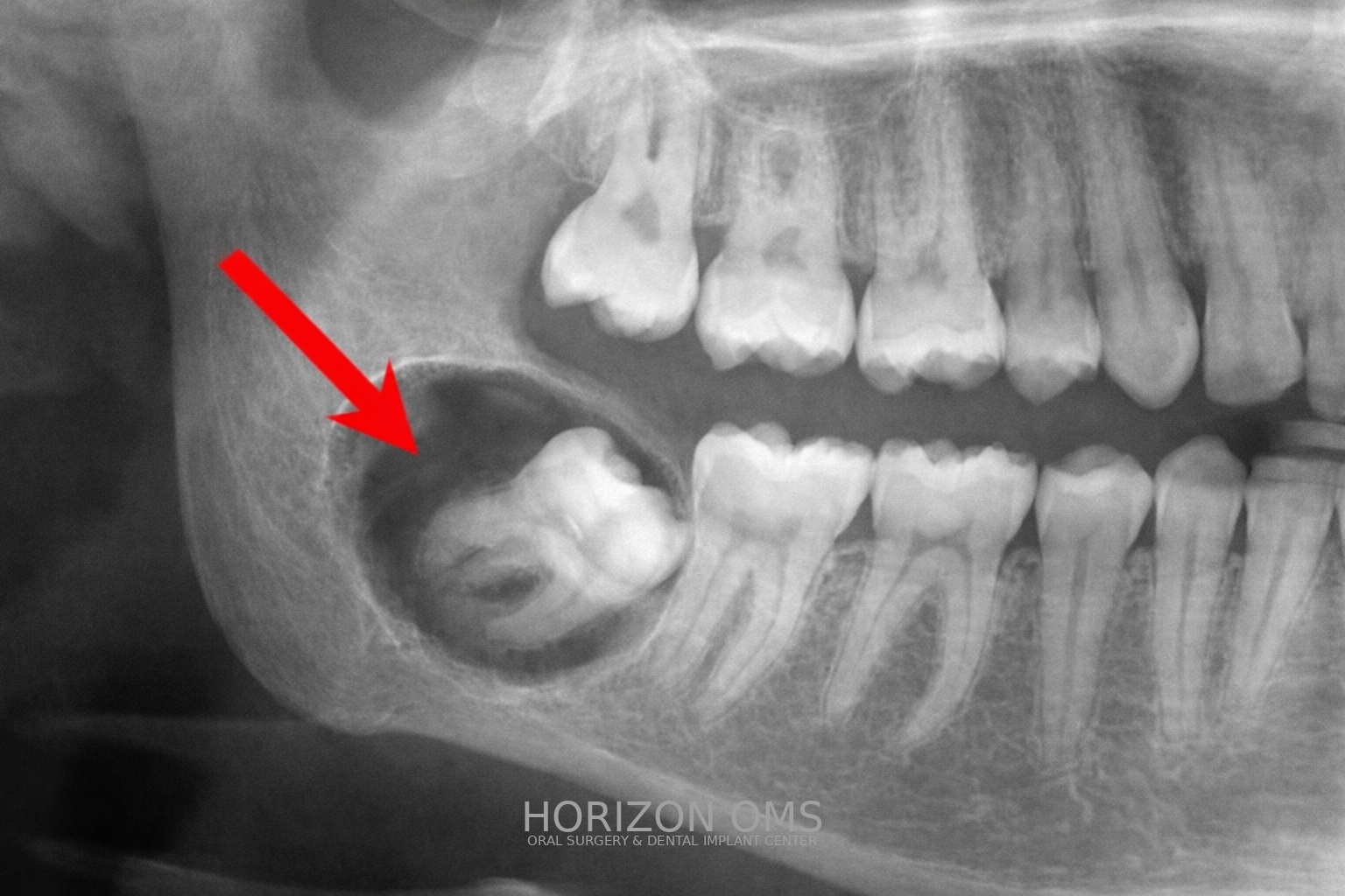
Evaluation of oral lesions
Not every spot or bump is serious — but anything that is new, changing, persistent, or painful deserves a proper exam. We evaluate the mouth, jaw, and surrounding tissues and recommend observation, imaging, or biopsy when appropriate.
Oral biopsy
A biopsy is a small surgical procedure used to obtain tissue for diagnosis. We perform soft tissue biopsies (for areas on the gum, cheek, tongue, or palate) and can also evaluate jaw (hard tissue) findings seen on imaging — often described as bony “lesions” or “cysts.” Depending on the finding, a biopsy may be performed under local anesthesia, with IV sedation, or in a hospital setting. We coordinate pathology review and communicate results and next steps.
Soft tissue vs. jaw (hard tissue) biopsies
- Soft tissue: gums, inner cheek, tongue, lip, or palate. Many can be completed in the office with local anesthesia.
- Jaw bone: lesions within the bone (commonly referred to as cysts). We typically review imaging (often CBCT when helpful) to plan a safe approach and determine whether an in-office procedure or sedation is best.
When a biopsy is considered
- A sore, ulcer, or lesion that does not heal
- Unexplained white or red lesions on the gum or tongue that last more than 2 weeks
- A growth or lump that is enlarging
- Persistent swelling, numbness, or unexplained pain
- Radiographic findings in the jaw that need clarification
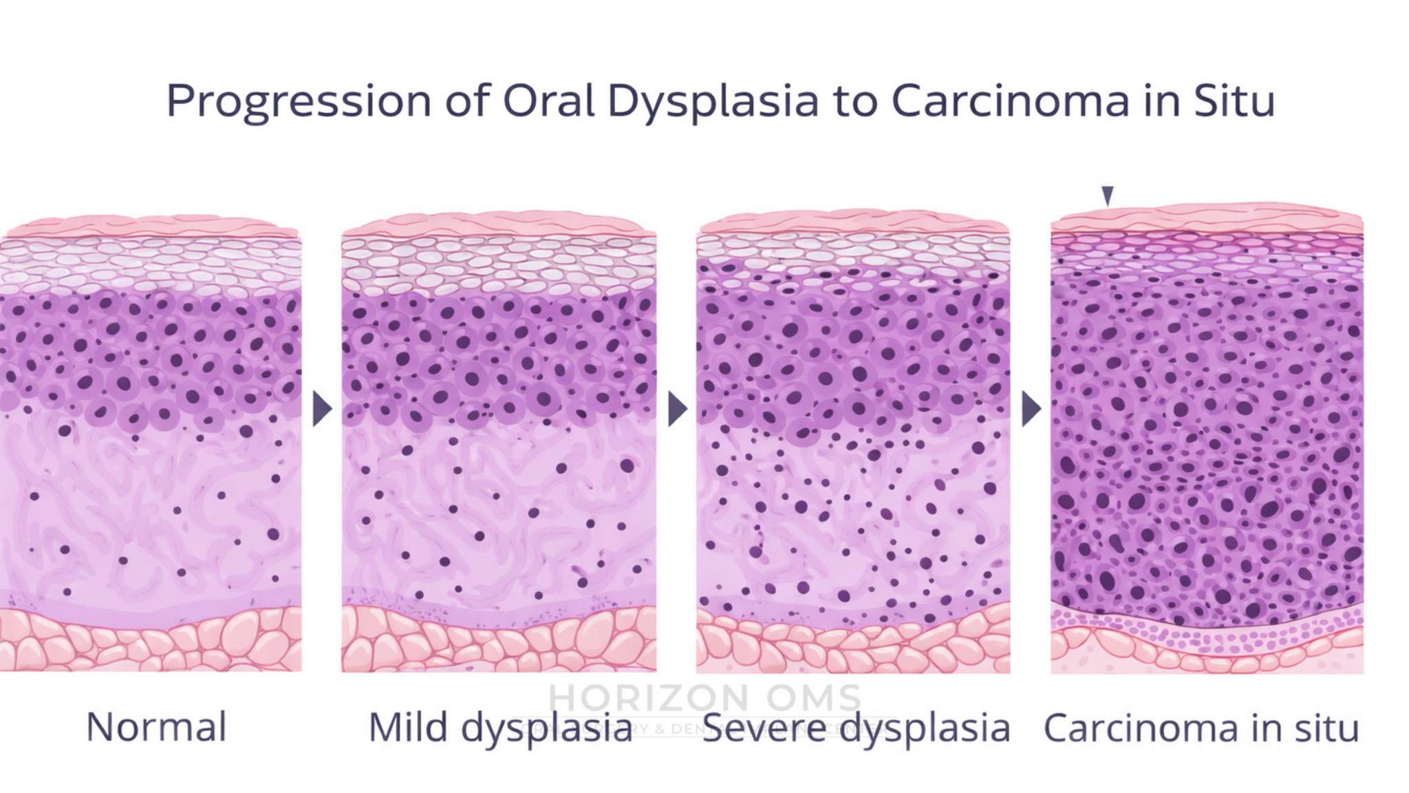
Oral epithelial dysplasia means there are abnormal changes in the surface lining cells. It is not a cancer diagnosis, but depending on the grade it can carry a risk of progression over time and may require monitoring, repeat biopsy, or removal/ablation in select cases. Read: Oral dysplasia & management (PDF).
Cases from Our Practice
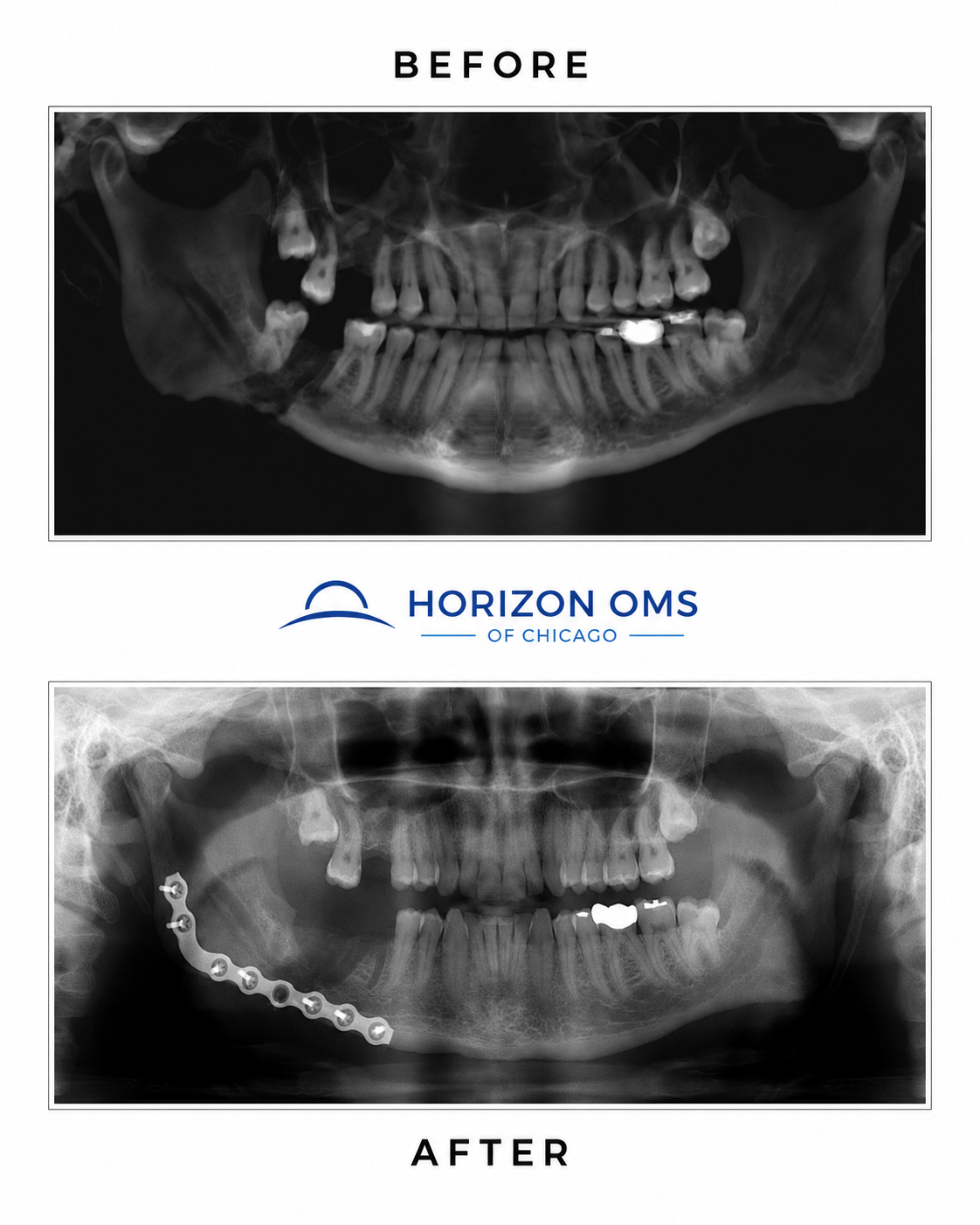
This patient was referred for management of a complex dental infection that had spread into the jawbone, known as osteomyelitis. The severity of the infection caused a fracture in his lower jaw, called a pathological fracture. Treatment involved surgical debridement to remove all infected and nonviable bone, followed by repair of the jaw with surgical hardware, including a reconstruction plate.
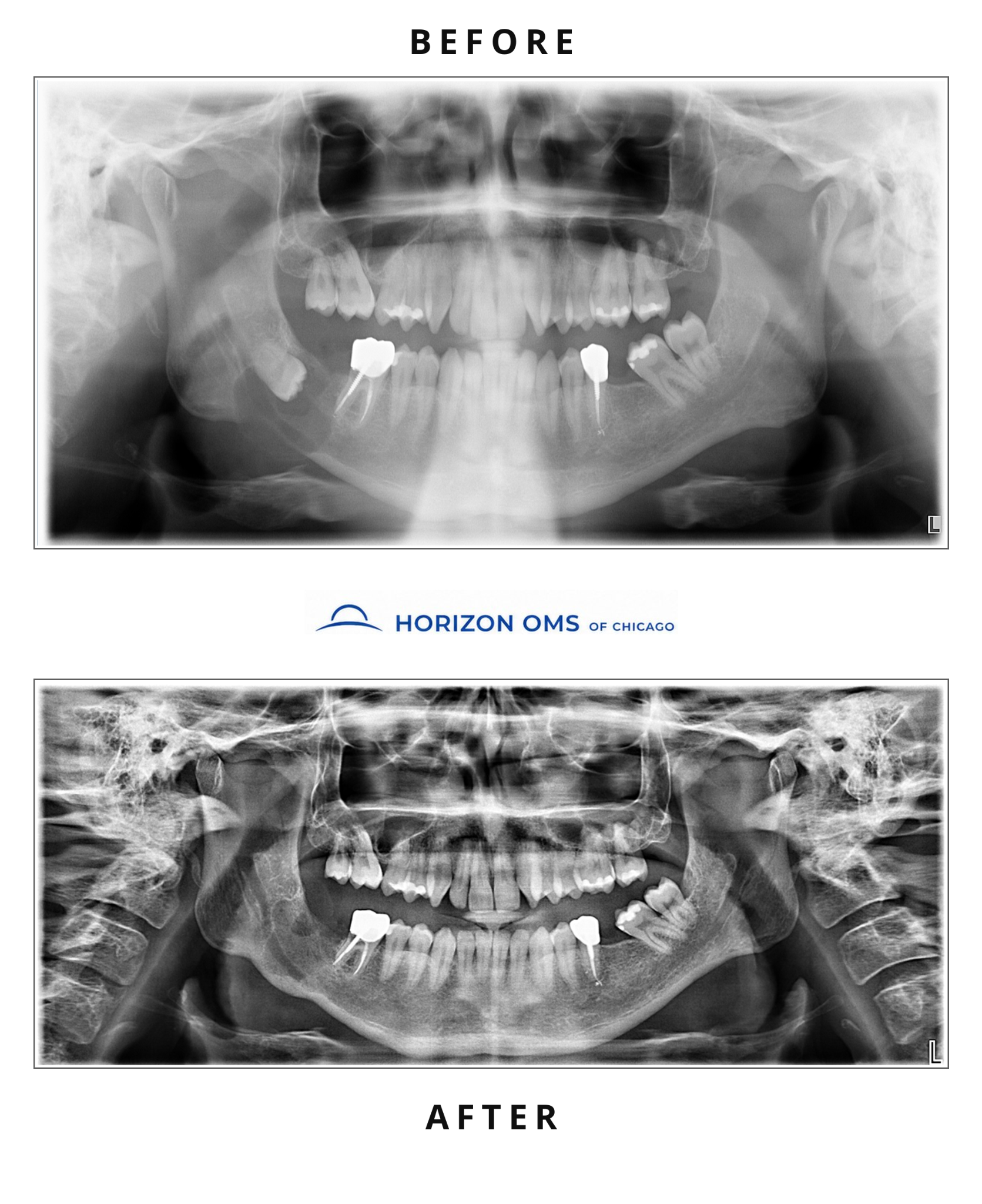
This patient was found to have a large cyst in the right jaw that was on the verge of causing a fracture because so much bone structure had been lost. The cyst was first decompressed, allowing it to shrink and decrease the risk of jaw fracture, followed by removal of the cyst with enucleation and curettage. This staged treatment avoided a jaw fracture as well as injury to the nerve that runs within the jawbone.
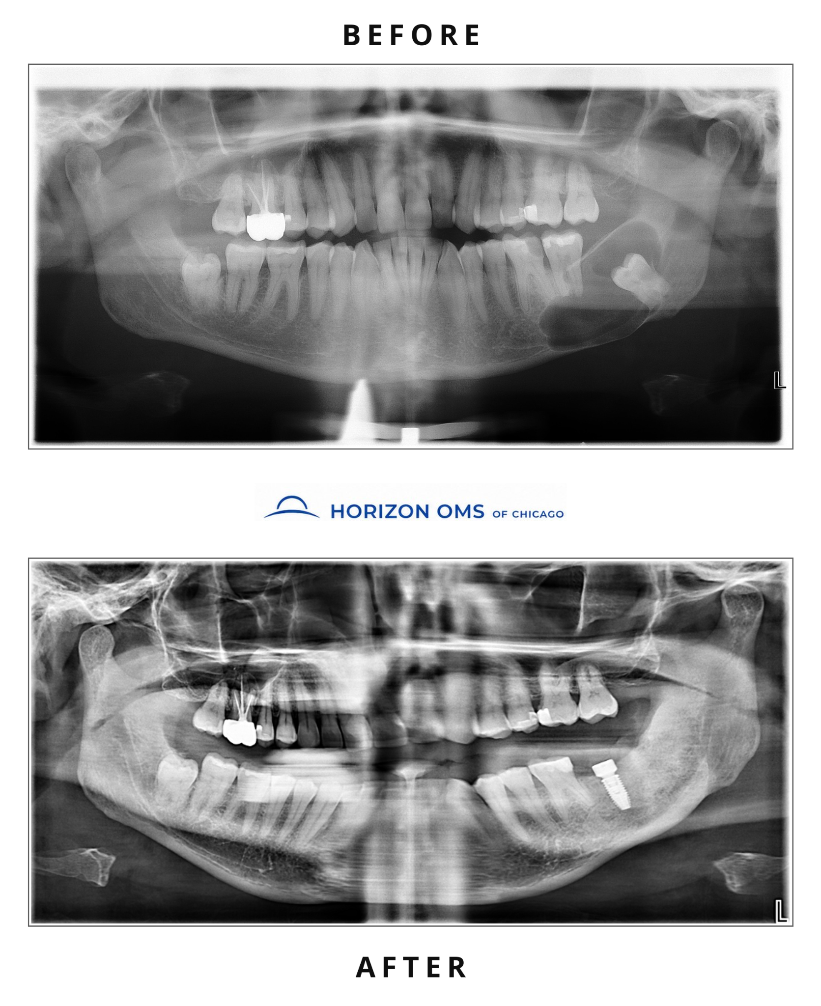
This patient was incidentally found to have an extremely large lesion in his left jaw during a routine exam by his dentist. The lesion was treated with decompression for several months to shrink it, followed by complete removal of the lesion and bone grafting. After healing, an implant was placed to restore the tooth that had been lost. Some cysts and jaw lesions can develop around impacted teeth, which is one reason timely evaluation and, when appropriate, wisdom teeth removal can help prevent pathology from developing.
Cases personally treated by Dr. Ali Al-Qudsi, board-certified oral and maxillofacial surgeon. Individual anatomy, healing, and results vary.